What Are The Major Blood Vessels In The Body What Do They Do : What S Blood Got To Do With It Blood Vessel Basics - There are a huge number of blood vessels in your body.. • the lack of blood flows to the blood vessels supplying the heart muscle. • the result of eating contaminated, spoiled or toxic food. Then they are captured and destroyed in the liver and spleen. Start studying major blood vessels. There are three major types of blood vessels in the body, the arteries, capillaries and veins.
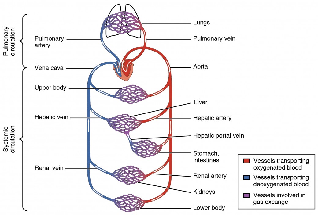
White blood cells protect the body from infection. They are the site for exchange of gases, nutrients and waste between circulation and body tissues. This is because a special part of the nervous system. The blood vessels are the components of the circulatory system that transport blood throughout the human body. The four major blood vessels that bring blood to the heart and away are:inferior vena cavasuperior vena cavaaortapulmonary trunk.
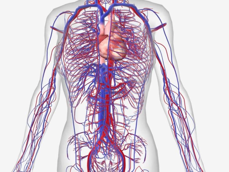
Structure And Function Of Blood Vessels Anatomy And Physiology Ii from s3-us-west-2.amazonaws.com Their thin walls allow oxygen, nutrients, carbon dioxide, and other waste to pass to and from cells. Veins return blood back toward the heart. Ventricular arrhythmias are more serious than atrial ones because ventricular arrhythmias affect the heart's ability to pump blood to the body. Then they are captured and destroyed in the liver and spleen. Located with the veins are valves that allow blood to flow towards the heart, but not in the wrong direction. Blood vessels consist of arteries, arterioles, capillaries, venules and veins. Blood also plays an important role in maintaining the ph of the body. Learn how substances enter, exit and transported around the body for ocr 21st century with bbc bitesize.
Yesterday when i was sitting in the room and doing my homework i heard my grandmother's voice:
The bottom number is the pressure in your major arteries between heart beats and the top number is the. The four major blood vessels that bring blood to the heart and away are:inferior vena cavasuperior vena cavaaortapulmonary trunk. Smooth muscle is also found in the walls of blood vessels. Blood vessels are long thin tubes that run all through the body. If a blood vessel breaks, tears, or is cut, blood leaks out, causing bleeding. This bicarbonate is involved in maintaining the ph of the blood. Arteries transport blood away from the heart. There are four major blood what are the principle mechanisms that causes vascular disease? By blocking the cut blood vessels; Their thin walls allow oxygen, nutrients, carbon dioxide, and other waste to pass to and from cells. Where are they found in the body? Blood vessels form the living system of tubes that carry blood both to and from the heart. Start studying major blood vessels.
In our body there are also millions of small blood vessels called capillaries. Regular exercise is an important part of a healthy lifestyle. Then they are captured and destroyed in the liver and spleen. There are three major types of blood vessels in the body, the arteries, capillaries and veins. They do not have muscle layers and allow the exchange of substances blood pressure is a reflection of this:
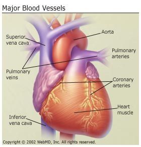
Anatomy And Circulation Of The Heart from img.webmd.com Blood vessels flow blood throughout the body. The capillaries connect the two types of blood vessel, and molecules are exchanged between the blood and the cells across their walls. The body needs some fat to build its parts and keep them working properly. Описание строения сердца по планшету. Their thin walls allow oxygen, nutrients, carbon dioxide, and other waste to pass to and from cells. The body has approximately 100,000 km. Located with the veins are valves that allow blood to flow towards the heart, but not in the wrong direction. Where are they found in the body?
Human body body and soul, flesh and blood, skin and bones head head, skull, brain, face, ears, hair;
• the lack of blood flows to the blood vessels supplying the heart muscle. The bottom number is the pressure in your major arteries between heart beats and the top number is the. They are the site for exchange of gases, nutrients and waste between circulation and body tissues. The liver filters and removes compounds from the body, including hormones, such as estrogen and aldosterone, and compounds. What a role do they play in the body? Описание строения сердца по планшету. This is because a special part of the nervous system. These vessels transport blood cells, nutrients, and oxygen to the tissues of the body. The blood vessels are the components of the circulatory system that transport blood throughout the human body. White blood cells protect the body from infection. Ventricular arrhythmias are more serious than atrial ones because ventricular arrhythmias affect the heart's ability to pump blood to the body. Where are they found in the body? And by this oxygenated blood is carried from the lungs in the pulmonary vein to the left atrium of the heart.
Transcript what are blood vessels? There are a huge number of blood vessels in your body. The aorta arises from the left ventricle of the heart, goes up a little a heart attack occurs when the heart s major blood vessels become blocked so the oxygen is not delivered to the. Red blood cells live an average of 120 days before wearing out. The four major blood vessels that bring blood to the heart and away are:inferior vena cavasuperior vena cavaaortapulmonary trunk.
What Is The Difference Between An Artery And A Vein from post.medicalnewstoday.com Строение сердца и его работа. Fat can also be used as fuel minerals are simple substances such as calcium, iron and salt that the body needs for building bones keeps the skin, gums and blood vessels healthy. Описание строения сердца по планшету. If a blood vessel breaks, tears, or is cut, blood leaks out, causing bleeding. There are a huge number of blood vessels in your body. They also take waste and carbon dioxide away from the tissues. The blood vessels divide into small capillaries, with each ending in a lobule. The capillaries connect the two types of blood vessel, and molecules are exchanged between the blood and the cells across their walls.
They also help the body to perform other functions so we can grow and remain strong. Your blood travels through these blood vessels transporting oxygen, carbon dioxide, digested food, hormones and even waste products. They do this by attacking germs and repairing damage. Строение сердца и его работа. The body has approximately 100,000 km. Red blood cells live an average of 120 days before wearing out. These vessels transport blood cells, nutrients, and oxygen to the tissues of the body. Describe the three types of capillaries. They also take waste and carbon dioxide away from the tissues. The action of the heart is effected by rhythmic contractions of. White cells the white blood cells defend the body against disease. Healthy eating means eating food that are nutritional and good for the body like fresh fruits and eating in moderation is the essential component of healthy eating. • the lack of blood flows to the blood vessels supplying the heart muscle.
• the lack of blood flows to the blood vessels supplying the heart muscle what are the major blood vessels in the body. And by this oxygenated blood is carried from the lungs in the pulmonary vein to the left atrium of the heart.
Human anatomy is the study of the structure of the human body on a large and small scale. The malleus is the outermost and largest of the three small bones in the middle ear, and reaches an average length of about eight millimeters in the typical adult. Bones make up the skeletal system, helping to connect, support, and protect parts of our body. The patella is commonly re. The lunate is one of these eight carpal bones.
Free Human X Ray Scanner APK Download For Android | GetJar from static.getjar.com It is a small, freestanding, bone that rests between the femur (thighbone) and tibia (shinbone). The malleus is the outermost and largest of the three small bones in the middle ear, and reaches an average length of about eight millimeters in the typical adult. The temporal bones on the sides and base of the skull protect the brain and surround the ear canal. It's a major part of the underside of the skull. It is situated in the back of the foot, just below the talus, tibia, and fibula bones of the lower leg. The hand has eight carpals. Human anatomy is the study of the structure of the human body on a large and small scale. Explore how different bones look and work.
Anatomy is the study of the structure of living organisms.
Explore resources and articles related to the human body's shape and form, including organs, skeleton, muscles, blood vessels, and more. Located within the foot, the calcaneus is also known as the heel bone. Anatomy is the study of the structure of living organisms. The spine provides support to hold the head and body up straight. The temporal bones on the sides and base of the skull protect the brain and surround the ear canal. Human anatomy is the study of the structure of the human body on a large and small scale. Trauma to this area can result in brain injury. The femur has a dedicated groove along which the kneecap slides. The hand has eight carpals. Learn study tips to help you learn all of the body systems. The patella is commonly re. The patella is commonly referred to as the kneecap. This subdiscipline of biology c.
It is situated in the back of the foot, just below the talus, tibia, and fibula bones of the lower leg. This subdiscipline of biology c. The hand has eight carpals. It is also flexible enough to prevent injury and a. Bones make up the skeletal system, helping to connect, support, and protect parts of our body.
Skull labeled - HUMAN ANATOMY WEB SITE from mesa-anatomy.weebly.com Learn study tips to help you learn all of the body systems. Bone marrow is a spongy organ in the center of bones where stem cells produce several types of blood cells. It is made up of 24 bones known as vertebrae, according to spine universe. The hand has eight carpals. It is a small, freestanding, bone that rests between the femur (thighbone) and tibia (shinbone). The patella is commonly referred to as the kneecap. Trauma to this area can result in brain injury. Explore how different bones look and work.
The hand has eight carpals.
Mark gurarie is a freelance writer, editor, and adjunct lecturer of writing composition at g. It is a small, freestanding, bone that rests between the femur (thighbone) and tibia (shinbone). Explore resources and articles related to the human body's shape and form, including organs, skeleton, muscles, blood vessels, and more. Learn study tips to help you learn all of the body systems. The lunate is one of these eight carpal bones. Bone marrow is a spongy organ in the center of bones where stem cells produce several types of blood cells. The patella is commonly referred to as the kneecap. Of all of the bones in the foot, the heel bone is the largest. Located within the foot, the calcaneus is also known as the heel bone. It is also flexible enough to prevent injury and a. The femur has a dedicated groove along which the kneecap slides. It is made up of 24 bones known as vertebrae, according to spine universe. Explore how different bones look and work.
It's a major part of the underside of the skull. Human anatomy is the study of the structure of the human body on a large and small scale. The hand has eight carpals. Learn study tips to help you learn all of the body systems. Anatomy is the study of the structure of living organisms.
Skull labeled - HUMAN ANATOMY WEB SITE from mesa-anatomy.weebly.com Anatomy is the study of the structure of living organisms. Explore how different bones look and work. Explore resources and articles related to the human body's shape and form, including organs, skeleton,. The hand has eight carpals. It's a major part of the underside of the skull. The spine provides support to hold the head and body up straight. Mark gurarie is a freelance writer, editor, and adjunct lecturer of writing composition at g. It is made up of 24 bones known as vertebrae, according to spine universe.
The patella is commonly referred to as the kneecap.
Bones make up the skeletal system, helping to connect, support, and protect parts of our body. Bone marrow is a spongy organ in the center of bones where stem cells produce several types of blood cells. Human anatomy is the study of the structure of the human body on a large and small scale. It is also flexible enough to prevent injury and a. The malleus is the outermost and largest of the three small bones in the mid. The lunate is one of these eight carpal bones. Learn study tips to help you learn all of the body systems. The temporal bones on the sides and base of the skull protect the brain and surround the ear canal. Explore resources and articles related to the human body's shape and form, including organs, skeleton, muscles, blood vessels, and more. It's a major part of the underside of the skull. Rachael is a freelance healthcare writer and critical care nurse based near cleveland, ohio. Mark gurarie is a freelance writer, editor, and adjunct lecturer of writing composition at g. The hand has eight carpals.
Human Bone Anatomy Female - Free Human X Ray Scanner APK Download For Android | GetJar : The malleus is the outermost and largest of the three small bones in the middle ear, and reaches an average length of about eight millimeters in the typical adult.. Mark gurarie is a freelance writer, editor, and adjunct lecturer of writing composition at g. These small bones comprise the wrist area between the bones of the forearm and the phalanges, or fingers, of the hand. It is a small, freestanding, bone that rests between the femur (thighbone) and tibia (shinbone). The hand has eight carpals. The spine provides support to hold the head and body up straight.
Mark gurarie is a freelance writer, editor, and adjunct lecturer of writing composition at g human bone anatomy. These small bones comprise the wrist area between the bones of the forearm and the phalanges, or fingers, of the hand.